rigid bronchoscopy

Rigid Bronchoscopy: 5 Essential Benefits of Advanced Airway Management

Introduction to Rigid Bronchoscopy

What is a Rigid Bronchoscopy?

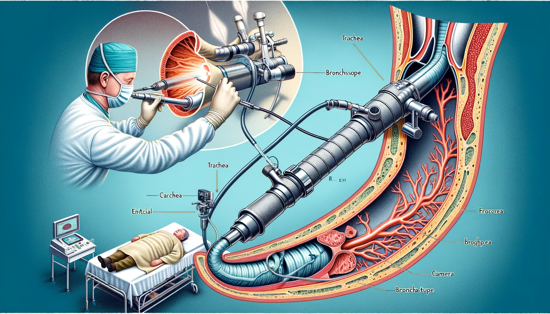
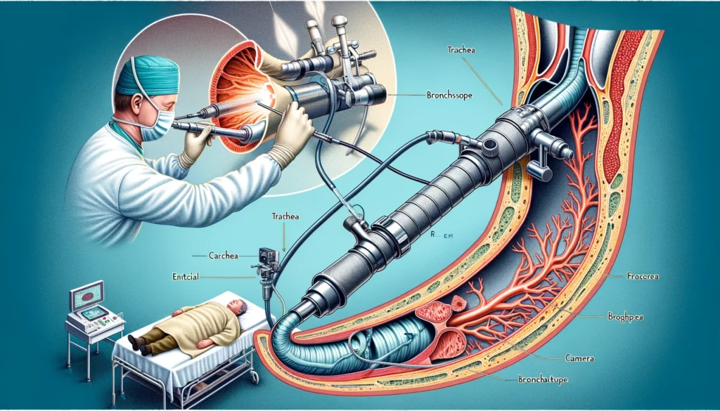
Rigid bronchoscopy is a specialized medical procedure that involves using a rigid bronchoscope to examine the lungs’ airways. This instrument is a straight, hollow tube equipped with a light and camera at its end, enabling doctors to visualize the trachea and bronchi directly. Unlike flexible bronchoscopy, which utilizes a more pliable scope, rigid bronchoscopy offers a more stable and larger working channel, making it particularly useful for certain diagnostic and therapeutic interventions. This procedure is essential for tasks requiring substantial airway control, such as removing foreign bodies, biopsy of suspicious lesions, stent placement, and managing central airway obstructions.
rigid bronchoscopy
rigid bronchoscopy

Historical Background

The roots of rigid bronchoscopy trace back to the late 19th century. In 1897, Dr. Gustav Killian performed the first bronchoscopy, marking a significant milestone in medical history. Over the decades, rigid bronchoscopy has evolved with advancements in technology and medical practices, enhancing its efficacy and safety. Initially, the procedure was primarily used for diagnostic purposes, such as inspecting the airways and identifying abnormalities. However, rigid bronchoscopy became a vital therapeutic tool as technology progressed. Its ability to provide a direct and unhindered view of the airways has made it indispensable in pulmonology and thoracic surgery.

Importance in Modern Medicine

In contemporary medical practice, rigid bronchoscopy is critical, especially in situations requiring immediate and precise intervention. Its significance is highlighted in emergency settings and critical care, where maintaining airway patency is crucial. Rigid bronchoscopy is often the preferred method for dealing with central airway obstructions, massive hemoptysis (bleeding), and complex therapeutic procedures requiring a stable and large working channel. The procedure’s ability to facilitate diagnostic and therapeutic interventions makes it a cornerstone in managing various respiratory conditions. As medical technology advances, rigid bronchoscopy remains a vital tool, contributing to improved patient outcomes and the advancement of respiratory care.

Indications for Rigid Bronchoscopy

Diagnostic Indications

Rigid bronchoscopy is frequently employed for diagnostic purposes in various clinical scenarios. It is particularly useful in obtaining tissue biopsies from the trachea and bronchi to diagnose lung cancers, infections, and other respiratory diseases. Additionally, it allows for direct visualization and assessment of airway abnormalities such as tumors, strictures, or congenital anomalies. In cases where imaging techniques such as CT scans or X-rays show ambiguous results, rigid bronchoscopy provides a more definitive evaluation, enabling clinicians to make accurate diagnoses and plan appropriate treatments.
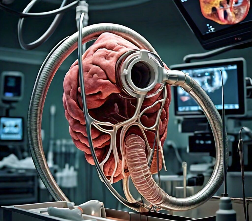
Rigid Bronchoscopy
Rigid Bronchoscopy

Therapeutic Indications

Beyond its diagnostic utility, rigid bronchoscopy plays a crucial therapeutic role. It is the procedure of choice for removing foreign bodies lodged in the airways, a common occurrence in pediatric cases. The rigid scope’s large lumen and stable structure make it ideal for manipulating and extracting objects that flexible bronchoscopes cannot handle as effectively. Furthermore, rigid bronchoscopy manages airway obstructions caused by tumors, benign growths, or scar tissues. It facilitates the placement of stents to keep the airways open and allows for laser therapy or mechanical debulking of obstructive lesions, providing immediate relief to patients with compromised airways.

Contraindications

Despite its benefits, rigid bronchoscopy is not suitable for every patient. Contraindications include severe hypoxia, uncontrolled bleeding disorders, and unstable cardiovascular conditions, as these factors can increase the risks associated with the procedure. Patients with significant cervical spine instability or severe tracheal stenosis may also be poor candidates due to the potential for airway trauma. A thorough pre-procedural assessment is essential to identify these contraindications and mitigate the risk of complications. Additionally, careful consideration must be given to the patient’s overall health status and the potential benefits and risks of performing the procedure.

Procedure Overview

Pre-procedural Preparation

Proper preparation is critical to the success and safety of a rigid bronchoscopy. Pre-procedural steps include a detailed medical history and physical examination to identify potential contraindications. Blood tests, including coagulation profiles, are performed to ensure the patient’s readiness for the procedure. Patients are typically required to fast for several hours before the bronchoscopy to reduce the risk of aspiration during anesthesia. The procedure is usually performed under general anesthesia, so pre-anesthetic evaluation and planning are crucial. After discussing the risks, benefits, and available options with the patient, informed consent is acquired.

Step-by-Step Procedure

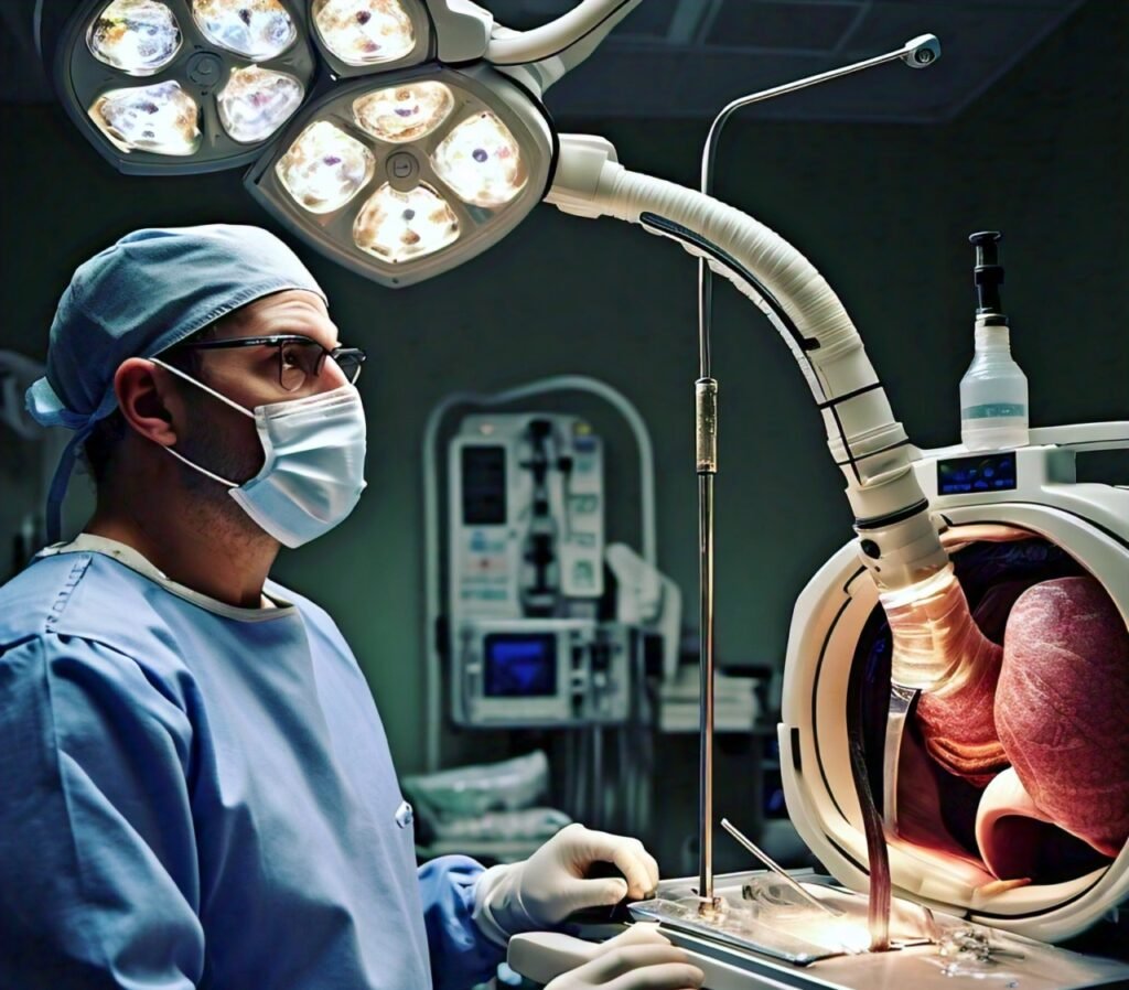
The rigid bronchoscopy procedure begins with the patient being positioned supine with the neck slightly extended to facilitate airway access. After the induction of general anesthesia, the rigid bronchoscope is carefully inserted through the mouth and advanced into the trachea. Continuous visualization is maintained using a camera attached to the scope. The bronchoscope allows for direct inspection of the tracheobronchial tree, and any abnormalities are noted. Depending on the indication, biopsies may be taken, foreign bodies removed, or therapeutic interventions such as stent placements or laser treatments performed. The procedure is meticulously monitored to ensure patient safety and effective airway management.

Post-procedural Care

Post-procedural care is vital to monitor and address any complications that may arise following a rigid bronchoscopy. Patients are observed in a recovery area until they regain full consciousness and their vital signs stabilize. Potential complications such as bleeding, infection, or respiratory distress are closely monitored. Pain management and supportive care, including oxygen therapy if needed, are provided based on the patient’s condition. Follow-up appointments are scheduled to assess the patient’s recovery and the success of the intervention. Patients are advised to report any persistent or worsening symptoms, such as difficulty breathing or chest pain, to their healthcare provider immediately.

Technological Advancements in Rigid Bronchoscopy

Instrumentation

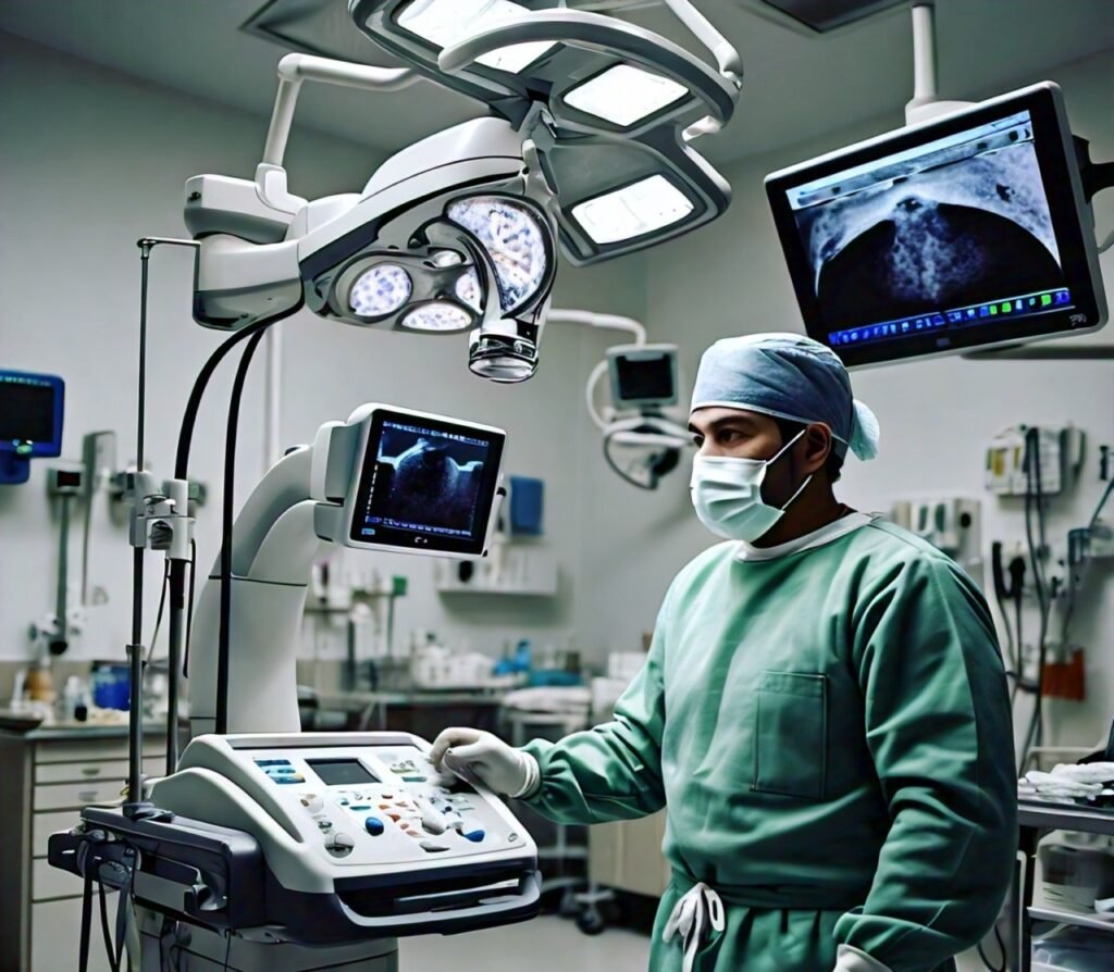
The field of rigid bronchoscopy has seen significant technological advancements, particularly in instrumentation. Modern rigid bronchoscopes are crafted from high-quality materials that enhance durability and flexibility, providing better maneuverability within the airways. These instruments have high-definition cameras that deliver clear and detailed visuals, essential for accurate diagnosis and effective treatment. Furthermore, various attachments, such as laser fibers, cryoprobes, and electrocautery tools, can be used with the rigid bronchoscope, expanding its therapeutic capabilities. These advancements have made rigid bronchoscopy safer and more efficient, allowing for a broader range of procedures to be performed with greater precision.

Rigid Bronchoscopy
Rigid Bronchoscopy

Imaging Enhancements

Imaging technology has dramatically improved the effectiveness of rigid bronchoscopy. High-definition video bronchoscopy allows for real-time, clear visualization of the airways, enhancing the ability to identify and treat abnormalities. Additionally, 3D imaging and virtual bronchoscopy provide a more comprehensive view of the bronchial tree, facilitating better planning and execution of complex procedures. Enhanced imaging modalities, like autofluorescence and narrow-band imaging, improve the detection of subtle lesions and early-stage cancers that might be missed with traditional white light bronchoscopy. These imaging enhancements are crucial in improving diagnostic accuracy and patient outcomes.

Integration with Other Modalities

Integrating rigid bronchoscopy with other medical modalities has further expanded its utility. Techniques such as endobronchial ultrasound (EBUS) and electromagnetic navigation bronchoscopy (ENB) can be combined with rigid bronchoscopy to provide a more detailed evaluation and treatment plan. EBUS, for example, allows for real-time ultrasound imaging of the airway walls and surrounding structures, facilitating the diagnosis and staging of lung cancer and other conditions. Similarly, ENB guides the bronchoscope to precise locations within the lung, aiding in the biopsy of peripheral nodules that are difficult to reach with traditional methods. This multidisciplinary approach enhances the diagnostic and therapeutic potential of rigid bronchoscopy.

Complications and Risks

Common Complications

Like any medical procedure, rigid bronchoscopy carries potential risks and complications. Common complications include bleeding, particularly if a biopsy or other invasive procedure is performed. Patients may also experience infection, due to the introduction of instruments into the airway. Other risks include damage to the teeth, lips, or throat, which can occur during the insertion of the rigid bronchoscope. Airway perforation, though rare, is a serious complication that requires immediate medical attention. Understanding these potential risks is essential for healthcare providers and patients to prepare for the procedure and recognize symptoms of complications early.

Managing Complications

Effective management of complications in rigid bronchoscopy is critical to ensure patient safety and successful outcomes. In cases of bleeding, applying pressure, using topical hemostatic agents, or performing electrocautery can help control the hemorrhage. If an infection develops, appropriate antibiotic therapy is initiated based on culture and sensitivity results. Careful technique during the procedure can minimize the risk of physical trauma, but if damage does occur, prompt repair or supportive measures, such as using soft protectors and carefully navigating the scope, are employed. Emergency surgical intervention may be necessary to repair the damage and prevent further complications in an airway perforation. Continuous monitoring and immediate response to adverse events are crucial in managing complications effectively.

Risk Mitigation Strategies

Several strategies can help mitigate the risks associated with rigid bronchoscopy. Pre-procedural assessment, including a thorough medical history and physical examination, helps identify patients at higher risk for complications. Advanced imaging techniques can aid in better planning and execution of the procedure, reducing the likelihood of complications. Ensuring that skilled and experienced bronchoscopists perform the procedure is another critical factor in minimizing risks. Additionally, adhering to sterile techniques and infection control protocols reduces the risk of postoperative infections. Educating patients about the procedure, potential risks, and signs of complications can also play a significant role in the early detection and management of adverse events. By implementing these risk mitigation strategies, the safety and efficacy of rigid bronchoscopy can be significantly enhanced.

Rigid Bronchoscopy vs. Flexible Bronchoscopy

Key Differences

Rigid bronchoscopy and flexible bronchoscopy are essential tools in pulmonology but differ significantly in structure, application, and technique. Rigid bronchoscopy utilizes a straight, inflexible metal tube, providing a wide, stable channel that allows for better control and the ability to handle larger instruments. In contrast, flexible bronchoscopy employs a bendable tube, which can navigate through more intricate and distal airways with greater ease. The choice between the two often depends on the specific clinical requirements, with rigid bronchoscopy being preferred for major interventions and flexible bronchoscopy for more detailed diagnostic evaluations.

Rigid Bronchoscopy
Rigid Bronchoscopy

Advantages and Disadvantages

Each type of bronchoscopy has its own set of advantages and disadvantages. Rigid bronchoscopy is highly effective for therapeutic procedures such as tumor removal, dilation of strictures, and foreign body extraction due to its larger working channel and superior airway control. However, it typically requires general anesthesia and is more invasive, leading to a higher potential for complications. Flexible bronchoscopy, on the other hand, can be performed under local anesthesia with sedation, is less invasive, and offers greater patient comfort. Its flexibility allows for detailed visualization of smaller airways but lacks the stability and wide channel provided by rigid bronchoscopes, limiting its utility in certain therapeutic interventions.

Clinical Scenarios for Each

The choice between rigid and flexible bronchoscopy is influenced by the clinical scenario. Rigid bronchoscopy is often the procedure of choice in emergency situations requiring immediate airway management, such as severe central airway obstructions, massive hemoptysis, or the retrieval of large foreign bodies. It is also preferred for procedures requiring the placement of stents or the use of laser therapy. Flexible bronchoscopy, with its superior maneuverability and patient tolerance, is more commonly used for diagnostic purposes, such as taking biopsies from distal airways, bronchoalveolar lavage, and detailed inspection of the bronchial tree. It is also useful for assessing diffuse lung diseases and diagnosing infections.

Training and Expertise

Training Requirements

Becoming proficient in rigid bronchoscopy requires extensive training and hands-on experience. Medical professionals typically undergo specialized training programs that cover the theoretical aspects of the procedure, the anatomy of the airways, and the handling of various bronchoscopic instruments. This foundational knowledge is crucial for understanding the complexities of the procedure and ensuring patient safety.

Skill Development

Skill development in rigid bronchoscopy involves a combination of supervised practice, simulation-based training, and real-life procedural experience. Trainees begin with simulations and cadaveric practice to develop hand-eye coordination, instrument handling, and procedural techniques. As they progress, they participate in supervised procedures on patients, gradually taking on more responsibility as they demonstrate competence. Continuous practice and exposure to various clinical scenarios are essential for developing the dexterity and decision-making skills necessary for proficient performance.

Certification and Continuing Education

Certification in rigid bronchoscopy often involves meeting specific educational and procedural requirements and passing a competency assessment. Organizations such as the American Board of Internal Medicine (ABIM) and other professional societies provide certification programs to ensure that practitioners meet the standards of care. Continuing education is equally important, as it keeps practitioners updated on the latest advancements, techniques, and best practices in the field. Regular participation in workshops, conferences, and training sessions is encouraged to maintain and enhance skills, ensuring high-quality patient care and adherence to evolving medical standards.

Future Directions and Research

Emerging Techniques

The field of rigid bronchoscopy is continuously evolving with the advent of emerging techniques aimed at improving procedural efficacy and patient outcomes. One notable advancement is the integration of endobronchial ultrasound (EBUS) with rigid bronchoscopy, which enhances the visualization of structures beyond the airway walls and facilitates more precise biopsies and interventions. Additionally, 3D imaging and virtual bronchoscopy advancements provide detailed pre-procedural planning and intraoperative navigation, leading to more accurate and safer procedures. Innovations in instrumentation, such as more refined and flexible rigid scopes and enhanced laser technologies, are also expanding the therapeutic capabilities of rigid bronchoscopy.

Research Trends

Current research in rigid bronchoscopy is focused on several key areas. One significant trend is the exploration of combined modality treatments, such as using rigid bronchoscopy in conjunction with photodynamic therapy (PDT) to treat central airway tumors. Studies are also investigating the use of advanced biomaterials for stent placement to improve biocompatibility and reduce complications like granulation tissue formation and stent migration. Another area of research is the optimization of anesthesia protocols to minimize risks and improve patient comfort during and after the procedure. Additionally, ongoing research examines the long-term outcomes of various rigid bronchoscopic interventions to refine guidelines and protocols for clinical practice.

Potential Developments

Several potential developments are poised to transform the practice of rigid bronchoscopy. Integrating artificial intelligence (AI) and machine learning algorithms into bronchoscopy could revolutionize diagnostic accuracy and procedural planning by providing real-time data analysis and decision support. AI could also enhance the interpretation of imaging and biopsy results, leading to earlier and more accurate diagnoses. Furthermore, developing new therapeutic agents and delivery mechanisms specifically designed for use with rigid bronchoscopy could expand its applications in treating complex airway diseases. Additionally, the miniaturization of tools and the enhancement of minimally invasive techniques promise to reduce procedure-related morbidity and expand the use of rigid bronchoscopy to a broader range of patients, including those with severe comorbidities who are currently considered high-risk.

Conclusion

Rigid bronchoscopy remains an indispensable tool in modern pulmonology, offering significant advantages for diagnostic and therapeutic purposes. Its ability to provide a stable, wide working channel makes it invaluable for procedures requiring substantial airway control, such as removing foreign bodies, managing central airway obstructions, and stent placements. Despite its invasiveness compared to flexible bronchoscopy, its precision and control in critical and complex interventions are unmatched. The evolution of rigid bronchoscopy is marked by advancements in technology and techniques, enhancing its efficacy and safety. Emerging innovations such as endobronchial ultrasound integration, 3D imaging, and virtual bronchoscopy are pushing the boundaries of what can be achieved. At the same time, ongoing research focuses on improving patient outcomes and expanding therapeutic applications.

FAQs

What is rigid bronchoscopy used for?

Rigid bronchoscopy is used to manage airway diseases for diagnostic and therapeutic purposes. It is commonly employed to remove foreign bodies, control massive bleeding in the airways, perform biopsies of central airway tumors, dilate strictures, and place stents to maintain open airways.

How is rigid bronchoscopy performed?

Rigid bronchoscopy is typically performed under general anesthesia. The patient is positioned appropriately, usually lying flat on the back, and a rigid bronchoscope is inserted through the mouth or nose into the trachea and bronchi.

What are the risks associated with rigid bronchoscopy?

Like any invasive procedure, rigid bronchoscopy carries certain risks. Common complications include bleeding, infection, and injury to the airways. Anaesthesia might sometimes have unfavourable effects.

How long does a rigid bronchoscopy procedure take?

The duration of a rigid bronchoscopy procedure can vary depending on the complexity of the case and the specific interventions required. Generally, the procedure itself takes between 30 minutes to an hour.

What is the difference between rigid and flexible bronchoscopy?

Rigid and flexible bronchoscopy differ primarily in the type of scope used and their respective applications. Rigid bronchoscopy uses a straight, inflexible metal tube, providing a stable and wide working channel suitable for major therapeutic interventions.

2 thoughts on “Rigid Bronchoscopy: 5 Essential Benefits of Advanced Airway Management

Leave a Reply

Your email address will not be published. Required fields are marked *